
Dr. Ketaki Marodkar
Assistant Professor,
Department of Anaesthesiology,
NKPSIMS, Nagpur
Introduced in 1941 and 1943, the Miller’s straight blade and Macintosh’s curved blade laryngoscopes respectively have been widely used for direct laryngoscopy and tracheal intubation. Although they have a good success rate of intubation, in 0.05% of surgical patients and in 0.13-0.35% of obstetric patients01, failed intubation occurs. Incidence of unsuspected difficult intubation is estimated to be still higher at 3%. One of the most important factors which contribute to difficult intubation is poor visualisation of glottis opening. Almost 30% of the anaesthesia-related deaths are induced by the complications of difficult airway management and more than 85% of all respiratory related complications cause brain injury or death. The pursuit to equip oneself with a better gadget for airway instrumentation led to introduction of various optical stylets and flexible fibrescopes but each of these had their own limitations.
The invention of CMOS (complementary metal oxide semiconductor) and large scale manufacture of video chips first led to development of video laryngoscopes (VL) to view the glottis so that trachea could be intubated. Video laryngoscopes have certainly made the task of securing the airway easier for experienced anaesthesiologists in difficult airway scenario, non-anesthesiologists during CPR and even in the pre-hospital settings for emergency care providers.
There are striking differences in usage and images of video laryngoscope and conventional laryngoscope, as described below:
- The VL system provides high-quality video images that are enlarged on the video monitor.
- If laryngeal manipulation is required to improve visualization of laryngeal structures, the intubating anaesthesiologist and the assistant can coordinate movements such as Sellicks manoeuvre as they observe simultaneously the image on the video monitor.
- With the video image projected from the distal end of the laryngoscope blade, laryngeal structures are kept in view as the ETT is passed through the oro-pharynx into the trachea.
- The anatomy of the blade is such that there is a steep curvature for alignment of oral, pharyngeal and laryngeal axes for viewing the glottis. The percentage of glottic opening (POGO) score is better than the Cormack and Lehane classification of laryngeal appearance.
- Shorter inter-incisor distance required for insertion of VL blade
Thus, with all these differences, the VL offers several important advantages over conventional laryngoscope which are as follows:
- Non-line-of-sight view and improved laryngeal visualization
- Less trauma to oral structures as less force used than during direct laryngoscopy
- Safer in cervical spine instability as minimal cervical spine movement
- Reduced hemodynamic stress response to laryngoscopy and intubation
- Easily portable
- Useful teaching tool
- Higher success rate in higher CL grade patients.
Conversely, the VL does have a few disadvantages too as follows:
- Passage of the ETT may be difficult despite good view and often stylet is needed
- Fogging and secretion may obscure the view
- Loss of depth perception
- Costly
- No single video laryngoscope is ideal
Given the advances in video technology, more reliable, powerful, and less expensive videolaryngoscopes are emerging. The most commonly used classification02 of VL is:
- Macintosh-modification
- Angulated blade
- Tube/guide channel
Macintosh-modification integrates video capability to the traditional Macintosh laryngoscope. The added advantage is the ability to perform both indirect; and in the case of video failure or secretions on the lens, direct laryngoscopy. This may also prove useful to educators in teaching novices of airway anatomy and ETI. These devices do not always require a stylet. Some examples are McGrath® Mac, Storz® V-Mac, and Storz® C-Mac.
Devices with an angulated blade incorporate a more angled curvature into the blade which markedly improves glottic visualization with minimal patient head flexion and neck extension. The endotracheal tube (ETT), which requires a matching pre-curved stylet, is carefully introduced and advanced “around the corner” until in monitor view, then maneuvered past the vocal cords at which time the stylet is removed and the ETT juts forth and downward. GlideScope® GVL, Cobalt, and Ranger, McGrath® Series 5 and Storz® C-Mac D-Blade are examples.
Tube/guide channel videolaryngoscopes use a guide channel to direct the preloaded ETT towards the glottis. Given the guide channel, a stylet is not necessary. All are designed for oral ETI using a standard ETT. They are highly portable given their small, directly attached video screens on the laryngoscope handle. Examples of tube/guide channel scopes are Pentax® AWS and Airtraq®.
Specific Video laryngoscopes and their salient features:
I] Glidescope03,04 Vascular and general surgeonJohn Allen Pacey designed the first video laryngoscope ‘Glidescope’ in 2001. It incorporates a high resolution digital camera, connected by a video cable to a high resolution LCD monitor. GlideScope owes its superior results to a combination of five key factors:
- The steep 60-degree angulation of its blade improves the view of the glottis by reducing the requirement for anterior displacement of the tongue.
- The CMOS APS digital camera is located at the point of angulation of the blade (rather than at the tip). This placement allows the operator to more effectively view the field in front of the camera.
- The video camera is recessed for protection from blood and secretions which might otherwise obstruct the view.
- The video camera has a relatively wide viewing angle of 50 degrees.
- The heated lens innovation helps to prevent fogging of the lens, which might otherwise obscure the view.
Tracheal intubation with the GlideScope can be facilitated by the use of the Verathon Stylet, a rigid stylet that is curved to follow the 60° angulation of the blade.
The GlideScope Ranger is a variant designed for use in pre-hospital airway management including air, land, and sea applications. This device weighs 1.5 pounds, and is waterproof as well as airworthy to 20,000 feet altitude.
The GlideScope Cobalt is a variant that has a reusable video camera with light-emitting core which has a disposable or single use external shell for prevention of cross infection. In August 2009, the world’s first tracheal intubation conducted with the assistance oftelemedicine technology was performed. During this demonstration, Dr. Sakles and the University of Arizona Telemedicine service guided physicians in a ruralhospital as they performed a tracheal intubation using the GlideScope.

II] Trueview Video Laryngoscope05
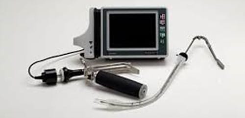
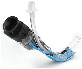
True view VL applies the optical principle of light refraction to provide a more anterior view of larynx and thus allow intubation to be performed under direct visualisation more frequently than is possible with a conventional laryngoscope. Truview PCDVL,with Truflex articulating stylet enables difficult oral and nasal intubations to be performed using a minimal amount of force and causing a reduced rate of patient side effects such as sore throat or soft tissue damage. It consists of reusable stainless steel blades, a viewtube, an oxygen insufflations port, a camera head that attaches to proximal part of view tube, a handle that provides the light source and a portable (5.5”battery powered) monitor [Figure 1, 2].
Distal end of its blade contains aprism with a 47 degree anterior view that refracts the line of vision and improves the C-L grade. The proximal lens magnifies the acquired image. An oxygen jet spray delivered via a unique injector, across the blade lenses during intubationprocedure serves to slow the rate of desaturation; prevent misting and remove secretions on the lensesthereby ensuring a clear visual picture throughout the entire intubation procedure.
III] Pentax Airway scope Video Laryngoscope06
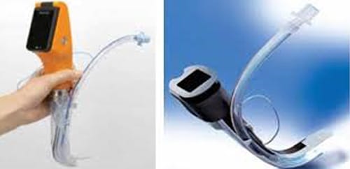
The compact, portable and water-resistant PENTAX Airway Scope video laryngoscope with high resolution colour images and a unique targeting system ensures accurate placement of ETT using compatible P Blades. The AWS offers a reduced degree of hemodynamic stimulation compared with the Macintosh laryngoscope, suggesting that tracheal intubation with the AWS is advantageous to prevent hypertension after laryngoscopy in high risk patients.
A video output port allows the user to connect the Airway Scope to a larger medical monitor or video recording device, making it possible to simultaneously show footage from the scope on a larger monitor or record for education or documentation.
With a 2.4 inch, 3 mega-pixel high-definition colour LCD screen providing vivid images of the larynx and glottis, and a cross-hair display on the screen, less experienced doctors will be able to reliably conduct tracheal intubation.
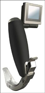
IV] King Vision Video Laryngoscope07
The King Vision Video laryngoscope is a two piece design. It is a high performance portable video laryngoscope which combines the convenience of a durable, reusable, video display with a disposable blade at an affordable price. The blades are disposable which eliminates the risk of cross contamination. Depending on the complexity of the airway, the King Vision has two blade types, both size 3 Macintosh but with difference in basic design:
- A standard blade that requires the use of a stylet to direct the ET tube
- A channeled blade where you can guide the ET tube with the blade loading it in a preformed channel on blade
The King Vision video laryngoscope provides a high quality image of the vocal cords while minimizing soft tissue manipulation.
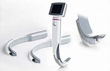
V] McGrath Video Laryngoscope08
- Easy to clean: The screen and handle are fully immersible for high-level disinfection, and compatible with the latest vaporized hydrogen peroxide sterilization systems.
- Slim Blade: The slim blade profile provides improved patient access. Blades are packaged sterile and are disposable.
- Portable: The compact, cable-free device is ready when you need it.
- Durable: Drop-tested to 2 meters, the core of the McGRATH® MAC enhanced direct laryngoscope is reinforced with a steel alloy chassis.
- Cost-effective: The surprisingly affordable system and blades make the McGRATH® MAC enhanced direct laryngoscope as accessible as your conventional laryngoscope, with the flexibility of video laryngoscopy.
VI] Airtraq Video Laryngoscope09
Airtraq is a novel single-use laryngoscope which provides glottis display without any deviation in the normal position of the oral, pharyngeal or the tracheal axes. Airtraq has an eyepiece optic, while the Airtraq Avant allows re-use of the optical system as well as external video monitoring and recording. The Airtraq blade is anatomically formed, with a battery-powered light source at the tip of the blade. The light bulb is designed to reach an operating temperature of 40 °C in order to suppress fogging of the optical system. The visual image is transmitted from a lens at the distal tip of the blade to the optical eyepiece (or monitor) via a series of prisms and mirrors.
Apart from the integrated optical system, the handle and blade of the Airtraq laryngoscope also contain a channel for the placement and insertion of the endotracheal tube. In patients having restricted neck motion or limited mouth opening (provided that it is greater than 3 cm) Airtraq offers the advantage of a better display. On the other hand the Airtraq possess certain disadvantages including the need of experience and the time demand for the operator to learn how to use them properly, the rapid deterioration of their display in the presence of a swelling or a secretion and the fact that they are rather complicated and expensive devices.
VII] Storz C-MAC Video laryngoscope10
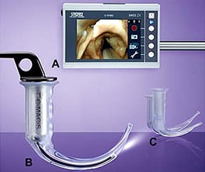
The C-MAC or Boedeker–Dörges videolaryngoscope is a modification of the V-MAC device. It includes an electronic module utilizing semiconductor chip technology and only consists of 3 parts, a laryngoscope, electronic module and separate 7-in (18 cm) monitor. The key innovation of C-MAC device is a completely portable setup that features an improved image quality. The C-MAC VL offers improved optics, field of view, interface for adjusting video quality, and easy recording of imaging. Moreover, the manufacturer provides the C-MAC device with a 2-inch pocket monitor attached to the handle. This portable device is specially developed for pre-hospital and in-hospital first aid. The device incorporates lithium-ion battery technology with at least 2 hours capacity and the pocket monitor can display a clear image under the strong light.
The C-MAC device can create continuous video recording or static pictures onto a removable secure digital card. The electronic module includes 2 buttons for photo and video capture.
There are 3 C-MAC reusable metal Macintosh blades (sizes 2–4) available for adult patients. The proximal flange of C-MAC reusable blade is significantly bigger than those of Glidescope and McGrath VL and is about 2.5 cm high at its base versus Glidescope VL, which is 1.5 cm and McGrath VL, which is 1.25 cm. This provides more space for manipulating the tracheal tube with the C-MAC device. In addition, the proximal shape and size of the C-MAC reusable blade make tube passage to the glottis more straightforward compared with that of the Glidescope or McGrath videolaryngoscope. The tip of the C-MAC reusable blade contains a 320×240 pixel complementary metal oxide semiconductor video chip and fog-resistant lens. The camera with the light source is located close to the tip of the blade and has an 80° view angle, allowing for a wide angle of viewing at the blade tip and a high-resolution color image on the monitor.
Future and Scope of VL:
Despite all the advantages offered, whether VL will replace traditional laryngoscopy is controversial. However, the American Society of Anesthesiologists (ASA) has already incorporated VL as an adjunct to ‘Alternative Difficult Intubation Approaches’ in their practice guidelines for management of the difficult airway. VL have quickly gained popularity as an intubation device in a variety of clinical scenarios and settings, as well as in the hands of airway experts and nonexperts. Their indirect view of the upper airway improves glottic visualization, including in suspected or encountered difficult intubation. Yet, more studies are needed to determine whether VL actually improves ETI success rates, intubation times, and first-attempt success rates; and thereby a potential replacement to traditional direct laryngoscopy.
References:
- Channa AB .Video laryngoscopes.. SJA 2011, Oct-Dec; 5(4):357-359.
- RV Chemsian, S Bhananker, and R Ramaiah. Videolaryngoscopy. Int J Crit Illn Inj Sci. 2014 Jan-Mar; 4(1): 35–41.
- Sharma D. Is GlideScope® the Best Way to Intubate?Anesthesiology 2010, Vol.113, 258-259
- C. Zaouter J. Calderon T. M. Hemmerling. Video laryngoscopy as a new standard of care. BJA:, Volume 114, Issue 2, February 2015, Pages 181–183 C. ZaouterC. ZaouterC. ZaouterC. ZaouterC. ZaouterC. ZaouterC. ZaouterC. Zaouter, J. Calderon, T. M. Hemmerling; Videolaryngoscopy as a new standard of care, BJA: British Journal of Anaesthesia, Volume 114, Issue 2, 1 February 2015, Pages 181–183,C. Zaouter, J. Calderon, T. M. Hemmerling; Videolaryngoscopy as a new standard of care, BJA: British Journal of Anaesthesia, Volume 114, Issue 2, 1 February 2015, Pages 181–183,C. Zaouter, J. Calderon, T. M. Hemmerling; Videolaryngoscopy as a new standard of care, BJA: British Journal of Anaesthesia, Volume 114, Issue 2, 1 February 2015, Pages 181–183,C. Zaouter, J. Calderon, T. M. Hemmerling; Videolaryngoscopy as a new standard of care, BJA: British Journal of Anaesthesia, Volume 114, Issue 2, 1 February 2015, Pages 181–183,C. Zaouter, J. Calderon, T. M. Hemmerling; Videolaryngoscopy as a new standard of care, BJA: British Journal of Anaesthesia, Volume 114, Issue 2, 1 February 2015, Pages 181–183,C. Zaouter, J. Calderon, T. M. Hemmerling; Videolaryngoscopy as a new standard of care, BJA: British Journal of Anaesthesia, Volume 114, Issue 2, 1 February 2015, Pages 181–183,C. Zaouter, J. Calderon, T. M. Hemmerling; Videolaryngoscopy as a new standard of care, BJA: British Journal of Anaesthesia, Volume 114, Issue 2, 1 February 2015, Pages 181–183,C. Zaouter, J. Calderon, T. M. Hemmerling; Videolaryngoscopy as a new standard of care, BJA: British Journal of Anaesthesia, Volume 114, Issue 2, 1 February 2015, Pages 181–183,C. Zaouter, J. Calderon, T. M. Hemmerling; Videolaryngoscopy as a new standard of care, BJA: British Journal of Anaesthesia, Volume 114, Issue 2, 1 February 2015, Pages 181–183,C. Zaouter, J. Calderon, T. M. Hemmerling; Videolaryngoscopy as a new standard of care, BJA: British Journal of Anaesthesia, Volume 114, Issue 2, 1 February 2015, Pages 181–183,C. Zaouter, J. Calderon, T. M. Hemmerling; Videolaryngoscopy as a new standard of care, BJA: British Journal of Anaesthesia, Volume 114, Issue 2, 1 February 2015, Pages 181–183,
- C. Zaouter, J. Calderon, T. M. Hemmerling; Videolaryngoscopy as a new standard of care, BJA: British Journal of Anaesthesia, Volume 114, Issue 2, 1 February 2015, PagesC. Zaouter, J. Calderon, T. M. Hemmerling; Videolaryngoscopy as a new standard of care, BJA: British Journal of Anaesthesia, Volume 114, Issue 2, 1 February 2015, Pages 181–183, Tiwari D, Chhabra S, Gehlaut P, Tiwari DP. Tracheal intubation using Truview PCDTM Video laryngoscope with Truflex Articulating Stylet in patients posted for lumbar spine surgery: A prospective study. Int J Med Res Prof. 2016; 2(1); 47-50.
- Nishikawa K, Matsuoka H, Saito S. Tracheal intubation with the PENTAX-AWS (airway scope) reduces changes of hemodynamic responses and bispectral index scores compared with the Macintosh laryngoscope J Neurosurg Anesthesiol. 2009 Oct; 21(4): 292-6.
- Kriege M, Aflen C, Noppens RR. Using King Vision Video laryngoscope with a channelled blade prolongs time for tracheal intubation in different training levels, compared to non channelled blade. Randomised Control Trial. PLoS One 2017 August 12(8):e0183382
- Ng, A.L.Hill, D.L.Williams, K.Lee, R.Segal. Randomized controlled trial comparing the McGrath videolaryngoscope with the C-MAC videolaryngoscope in intubating adult patients with potential difficult airways. BJA September 2012, Volume 109, Issue 3, Pages 439-443.
- Saracoglu KT, Eti Z, Gogus FY. Airtraq optical laryngoscope: advantages and disadvantages. Middle East J Anaesthesiol. 2013 Jun; 22(2): 135-41
- Fu-Shan Xue, Hui-Xian Li, Ya-Yang Liu, and Gui-Zhen Yang. Current evidence for the use of C-MAC videolaryngoscope in adult airway management: a review of the literature. Ther Clin Risk Manag. 2017; 13: 831–841.


